
Combining Noise-to-Image and Image-to-Image GANs: A Breakthrough in Brain MR Image Augmentation
Combining Noise-to-Image and Image-to-Image GANs: Brain MR Image Augmentation for Tumor Detection
The paper proposes a novel two-step data augmentation (DA) framework for brain tumor detection that combines noise-to-image (PGGANs) and image-to-image (MUNIT/SimGAN) GANs. By generating realistic 256x256 MR images and subsequently refining them, the method achieves a state-of-the-art sensitivity of 97.48% in tumor classification using ResNet-50.
TL;DR
To tackle the chronic shortage of annotated medical data, this study introduces a two-step GAN pipeline. By first generating whole-brain MRIs from noise using PGGANs and then refining them via MUNIT/SimGAN, the researchers pushed tumor detection sensitivity to a remarkable 97.48%. This work proves that synthetic data can effectively "fill the gaps" in real pathological distributions.
Problem & Motivation: The "Small Data" Wall in Radiology
Deep learning thrives on data, but medical imaging lives in a world of scarcity. While classic Data Augmentation (DA)—like flipping or rotating images—helps, it doesn't add new pathological information; it simply reshuffles existing pixels.
The authors identified a critical gap:
- Noise-to-Image GANs (like PGGAN) are great at creating diverse global structures but often fail at fine-grained medical textures.
- Image-to-Image GANs (like MUNIT) are excellent at refining textures but require an input "template" to start with.
By combining them, the authors aimed to create "infinite" new, realistic brain tumor samples that are both structurally diverse and texturally accurate.
Methodology: The Two-Step Generative Dance
The proposed framework treats image generation as a multi-stage refinement process rather than a single-shot attempt.
- Stage 1: Structural Generation (PGGANs)
Starting from random Gaussian noise, the PGGAN grows layers progressively (from 4x4 up to 256x256). This allows the model to learn the stable "macro" structure of a human brain before worrying about the "micro" details of a tumor. - Stage 2: Texture Refinement (MUNIT/SimGAN)
The 256x256 output from Stage 1 often carries "artifacts" or unrealistic intensities. The authors use MUNIT (which combines GANs and VAEs) to translate these synthetic images into the "style" of real MRIs, ensuring the tumor boundaries and tissue textures are clinically plausible.
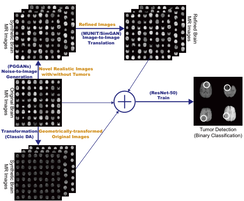 Figure 1: The two-step pipeline: PGGAN generates the base image, MUNIT refines it, and a ResNet-50 classifier performs the final diagnostic task.
Experiments and Results: Shifting the Manifold
The authors didn't just look at accuracy; they focused on Sensitivity—the most crucial metric in medicine, as missing a tumor (False Negative) is far more dangerous than a false alarm.
Key Findings:
- Sensitivity Boost: Combining classic DA with MUNIT-refined GAN images increased sensitivity from 93.67% to 97.48%.
- Visual Turing Test: An expert physician struggled to distinguish PGGAN-generated images from real ones, though refinement sometimes introduced minor artifacts that were human-detectable but actually helped the AI learn more robust features.
- The Power of t-SNE: As shown in the visualization below, GAN-generated images (especially MUNIT-refined ones) cover a wider and more distinct area of the feature space compared to simple geometric transformations.
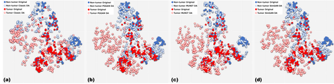 Figure 2: t-SNE plots showing that GAN-generated images (c & d) better fill the distribution gaps compared to classic DA (a).
Critical Analysis & Conclusion
This paper is a masterclass in Ensemble Generation. By decoupling the "what" (structure) from the "how" (texture), the authors bypassed the stability issues usually associated with training high-resolution medical GANs.
Limitations:
- The process requires a "discarding" step for "weird-looking" images, suggesting the GAN still lacks 100% reliability.
- It relies on 2D slices. In a clinical setting, tumors are 3D volumes.
Future Outlook: The authors suggest that this approach could be adapted for data anonymization, allowing hospitals to share "synthetic patients" for research without violating privacy laws. As we move toward 2026, the integration of such "two-step" generative models into standard medical AI pipelines seems inevitable.